High-resolution thyroid ultrasound in Nairobi
Expert thyroid imaging for accurate diagnosis and personalised care. Every scan is performed and reported by a consultant with dedicated thyroid expertise — using high-frequency ultrasound to assess your gland, grade any nodules with ACR TI-RADS, and give you clear answers at the same visit.
What is a thyroid ultrasound?
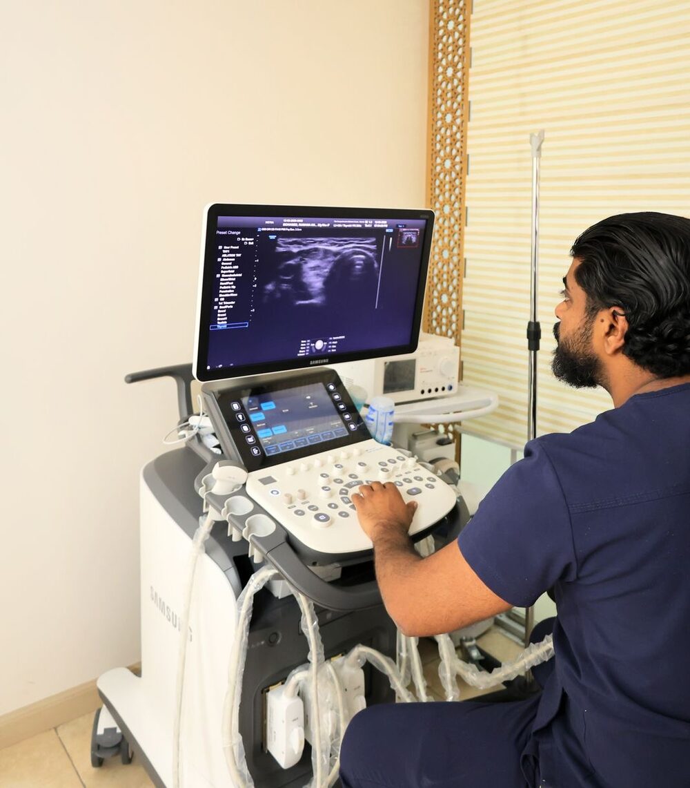
A thyroid ultrasound is a painless, radiation-free scan that uses high-frequency sound waves to produce detailed, real-time images of the thyroid — the butterfly-shaped gland at the front of your neck. A small handheld probe is moved gently over the skin; there are no needles, no injections and no ionising radiation, so it is completely safe to repeat as often as your care needs.
It is the single most important test for looking at the thyroid. Ultrasound shows the size and texture of the gland, maps any nodules in fine detail, assesses blood flow, and checks the lymph nodes of the neck — information that blood tests alone can never provide. For that reason it sits at the centre of almost every thyroid assessment we do.
Worth knowing
Blood tests tell us how the thyroid is working; ultrasound tells us how it is built. The two answer different questions — which is why a full assessment nearly always needs both.
Why you might need a thyroid ultrasound
You might be referred for a thyroid ultrasound, or arrange one yourself, for any of the following reasons.
A lump or neck swelling
To examine a swelling you can see or feel at the front of the neck, or a lump found by a doctor during examination.
A nodule found on another scan
To characterise a thyroid nodule discovered incidentally on a CT, MRI or carotid Doppler done for another reason.
Abnormal thyroid blood tests
To look at the gland when TSH, Free T4 or antibodies are abnormal — an overactive, underactive or autoimmune thyroid.
Symptoms in the neck
For difficulty swallowing, a sensation of fullness or pressure, hoarseness, or a chronic cough with no clear cause.
Monitoring a known nodule
To re-measure a nodule already under surveillance and compare it accurately against previous scans.
A family or personal history
For a family history of thyroid disease or cancer, or previous radiation to the head or neck, especially in childhood.
To guide a procedure
As the live imaging that guides an FNA or core biopsy, a cyst aspiration, or radiofrequency ablation (RFA).
Reassurance and a baseline
For a clear, documented baseline of a healthy thyroid — so any future change can be measured against it with confidence.
What happens during a thyroid ultrasound?
The scan is straightforward, comfortable and usually finished within about 15–20 minutes. Here is exactly what to expect.
Settle & recline
You lie back comfortably with a soft pillow under your shoulders and your neck gently extended. No fasting or preparation is needed.
A little gel
A water-based gel is applied to the skin of your neck so the probe glides smoothly. It's warmed where possible and wipes off easily afterwards.
The scan itself
The specialist moves the probe over your neck, viewing the gland live on screen. You'll feel light, painless pressure — nothing more.
Careful measurement
Each lobe, any nodule and the neck lymph nodes are measured in three dimensions and assessed for texture, margins and blood flow.
Same-visit explanation
Because a specialist is performing the scan, you discuss the findings there and then — not days later by letter.
Next steps agreed
Together we decide what, if anything, follows: reassurance, a repeat scan interval, blood tests, a biopsy, or treatment.
Why specialist thyroid ultrasound matters
Ultrasound is highly operator-dependent: the images are only as good as the person acquiring and interpreting them. A thyroid scan performed and read by a specialist is a fundamentally different test from a quick, delegated look.
- The scan is performed by the same specialist who interprets it — nothing is lost in translation between sonographer and reporter.
- Nodules are graded live with ACR TI-RADS, so biopsy decisions follow evidence rather than habit.
- Subtle but important features — micro-calcifications, irregular margins, abnormal lymph nodes — are actively looked for, not missed.
- Findings are explained to you at the visit, with time for your questions.
- The report is structured and reproducible, so future scans compare like with like.
- Where a biopsy or RFA is needed, it can often be discussed — and arranged — in the same clinic.
A confident, accurate scan spares many patients an unnecessary biopsy — and makes sure the few who genuinely need one are identified without delay.
Conditions we can diagnose and monitor
A single high-resolution scan gives us a detailed picture of a wide range of thyroid conditions — to diagnose them, grade their risk, and monitor them over time.
Thyroid nodules
Sized, characterised and graded with TI-RADS — the great majority proving benign.
Learn more →
Goitre
A diffusely or nodular enlarged thyroid — volume measured and the cause investigated.
Learn more →
Hyperthyroidism & Graves'
An overactive thyroid; ultrasound shows the gland's blood flow and rules out a toxic nodule.
Learn more →
Hypothyroidism & Hashimoto's
Autoimmune thyroid disease has a characteristic ultrasound pattern that supports the diagnosis.
Learn more →
Thyroid cysts
Fluid-filled nodules, almost always benign — and often treatable by simple aspiration.
Learn more →
Suspicion of thyroid cancer
Suspicious features and abnormal neck lymph nodes are identified early and acted on promptly.
Learn more →
Understanding your thyroid ultrasound report
A good ultrasound report is more than a picture — it's a structured record that any clinician can act on. Here is what yours describes.
What the scan measures
- Thyroid size and total gland volume
- Each lobe and the isthmus
- The number, size and position of any nodules
- Composition — solid, cystic or mixed
- Echogenicity, shape and margins
- Calcifications and other risk features
- Blood-flow (Doppler) pattern
- The cervical lymph nodes of the neck
Reading the language of the report
- Echogenicity — How bright or dark a nodule appears. Very dark ('markedly hypoechoic') nodules carry a little more risk and are graded accordingly.
- Composition — Whether a nodule is solid, fluid-filled (cystic) or a mix. Purely cystic nodules are almost always benign.
- Margins — The edge of a nodule. Smooth, well-defined margins are reassuring; irregular or lobulated margins raise the score.
- Calcifications — Tiny calcium deposits. Fine 'micro-calcifications' are a risk feature; coarse or rim calcification usually is not.
- Vascularity — The blood-flow pattern seen on Doppler, which helps distinguish an overactive nodule and adds context to the grading.
- TI-RADS score — The overall risk category, from TR1 to TR5, that ties all the features together into a clear recommendation.
Understanding ACR TI-RADS
ACR TI-RADS (Thyroid Imaging Reporting and Data System) scores a nodule's ultrasound features — composition, echogenicity, shape, margins and calcifications — and places it in one of five categories. The category, together with size, tells us whether a biopsy is needed at all.
TR1 — Benign
No suspicious features. No biopsy needed.
TR2 — Not suspicious
Benign-pattern nodule. No biopsy needed.
TR3 — Mildly suspicious
Biopsy considered from about 2.5 cm; ultrasound follow-up from 1.5 cm.
TR4 — Moderately suspicious
Biopsy considered from about 1.5 cm; follow-up from 1 cm.
TR5 — Highly suspicious
Biopsy considered from about 1 cm; close surveillance even when smaller.
The size thresholds are guideline figures — your overall picture decides. Graded properly, most nodules never need a needle at all. Specialist interpretation of the scan is what makes the grading trustworthy.
When is a thyroid biopsy needed?
A biopsy answers the one question a scan can't: what the cells themselves are doing. But most nodules never need one — it is recommended only when the ultrasound picture calls for it.
A biopsy is considered when
- The TI-RADS grade and nodule size cross the evidence-based threshold
- A nodule has grown meaningfully since a previous scan
- Suspicious features are present — irregular margins, micro-calcifications, a very dark solid nodule
- Abnormal-looking neck lymph nodes are seen alongside a nodule
- There is a family history of thyroid cancer or previous neck radiation
When it isn't needed
If your nodule doesn't cross these thresholds, the right answer is reassurance and a sensible follow-up interval — not a needle. Grading every nodule with TI-RADS is precisely what spares most people an unnecessary biopsy.
When a biopsy is needed it is quick, image-guided and done in clinic under local anaesthetic if required — with a small plaster and a return to your normal day.
Ultrasound-guided procedures
Live ultrasound doesn't just diagnose — it guides treatment with millimetre precision. Every image-guided procedure at the clinic is performed under the same high-resolution imaging.
Fine-needle aspiration (FNAC)
A very fine needle samples cells from a nodule under live guidance — quick, minimally uncomfortable, reported on the Bethesda system.
Learn more →
Core needle biopsy (CNB)
Takes a fine cylinder of tissue when FNA is repeatedly inconclusive — done under local anaesthetic with ultrasound guidance.
Learn more →
Cyst aspiration
Fluid-filled nodules can be drained in clinic under guidance; cysts that refill respond well to ablation.
Learn more →
Radiofrequency ablation (RFA)
Benign, symptomatic nodules shrunk from within — ultrasound-guided, no incision, no scar, home the same day.
Learn more →
Why high-quality ultrasound reporting matters
The value of a thyroid ultrasound lives or dies on the quality of its reporting. A vague report — 'nodule seen, clinically correlate' — leaves you and your doctor exactly where you started. A structured report gives clear measurements, a TI-RADS grade, and an explicit recommendation.
We report every scan to international standards: the size of each nodule in three dimensions, its features described systematically, its ACR TI-RADS category, and a plain recommendation for what should happen next. That structure is what makes a scan genuinely useful — and what makes the next scan a fair comparison.
What a good report gives you
- Measurable — Precise three-dimensional measurements, so growth or shrinkage is tracked accurately over time.
- Graded — An explicit ACR TI-RADS category for every nodule — not just a description.
- Actionable — A clear recommendation: reassure, re-scan at a set interval, biopsy, or treat.
- Reproducible — A consistent format so your next scan — here or elsewhere — compares like with like.
Thyroid ultrasound — your questions answered
No. It is a completely painless, radiation-free scan. A small probe is moved gently over the skin of your neck with a little gel — most people feel only light pressure, and it typically takes about 15–20 minutes.
No preparation is needed. You don't have to fast, stop any medication or change your routine. It helps to wear a top with an open or low neckline so the neck is easy to access.
No. You're welcome to arrange a thyroid ultrasound directly. If you do have a referral, previous scans or blood results, bring them along — they help us compare and interpret accurately.
It is very safe and uses no ionising radiation at all. Ultrasound uses harmless sound waves, so it's safe in pregnancy and can be repeated as often as your care requires.
The scan itself takes about 15–20 minutes. Because a specialist performs it, you discuss the findings at the same visit rather than waiting days for a report.
Usually not. We grade every nodule with ACR TI-RADS, and a biopsy is recommended only when a nodule's features and size cross evidence-based thresholds. Most nodules never need one.
It's an international scoring system that rates a nodule's ultrasound features and places it in one of five risk categories (TR1–TR5). The category, with the nodule's size, decides whether a biopsy is needed — so decisions follow evidence rather than habit.
It can't give a definitive answer on its own, but it's very good at sorting risk. Reassuring features suggest a nodule is benign; suspicious features prompt a biopsy, which confirms the diagnosis. Only around 5–10% of nodules prove cancerous.
Yes, completely safely. There is no radiation, so ultrasound is the ideal way to assess the thyroid during pregnancy. Any treatment that isn't urgent is simply planned for after delivery.
They answer different questions. Blood tests show how the thyroid is working (its hormone levels); ultrasound shows how it is built (its structure and any nodules). A full assessment usually needs both.
Often, yes. Because the specialist performs the scan, an FNA, cyst aspiration or a treatment discussion can frequently happen at the same visit where it's appropriate — sparing you repeat trips.
It depends on the findings. A benign nodule on surveillance is typically re-scanned every 12–24 months, spacing out as stability is confirmed. Your specialist sets the right interval for you.
Yes. Bring or send your previous images and report and we'll review them, re-scan where needed, and give you a clear structured opinion and plan.
Yes. Patients travel to us from across East Africa. Send your details or previous scans first on WhatsApp; we'll advise on what to bring and can often combine your scan, review and any treatment into a single short visit.
Imaging you can build decisions on
- Consultant endocrinologist with dedicated thyroidology expertise
- High-frequency ultrasound, performed and reported by the specialist
- ACR TI-RADS grading on every nodule, aligned with ATA, ETA and BTA guidance
- Same-visit findings, explained to you in plain language
- Image-guided FNA, core biopsy, cyst aspiration and RFA under one roof
- Structured, reproducible reports your referring doctor can act on
How your scan works
Scan
A thorough, specialist-performed ultrasound of the whole gland and neck.
Grade
Every nodule graded with ACR TI-RADS — evidence, not guesswork.
Explain
Your findings, discussed with you at the same visit.
Plan
A clear next step — reassurance, follow-up, biopsy or treatment.
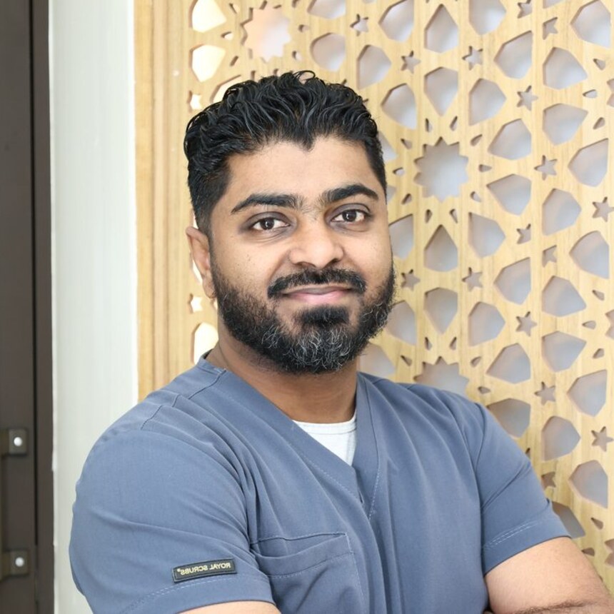
Dr. Ebrahim Yusuf Ebrahim
Consultant Physician · Interventional Endocrinologist & Diabetologist
One of the first doctors in Kenya to bring minimally invasive thermal ablation — microwave and radiofrequency — to the treatment of thyroid nodules and goitre, sparing patients from surgery. Every thyroid ultrasound at the clinic is performed and reported by him personally — so the scan and its interpretation are never separated.
- MMed, Internal Medicine — University of Nairobi
- PG Diploma, Endocrinology & Diabetes — Royal College of Physicians, Ireland
- Fellowship, Endocrinology & Diabetes — University of Alexandria, Egypt
- Certificate in Interventional Thyroidology — Mumbai, India
- Special focus: thermal ablation (RFA & microwave) for thyroid nodules
Refer a patient for thyroid imaging
We welcome imaging referrals from ENT surgeons, endocrinologists, radiologists and family physicians across the region. You receive a structured, consultant-reported scan — and your patient comes back to you with a clear plan.
Consultant-reported imaging
Every scan is performed and reported by the specialist, with three-dimensional measurements, ACR TI-RADS grading and an explicit recommendation.
One-stop assessment
Where indicated, ultrasound, FNA or core biopsy and treatment discussion can happen at the same visit — sparing your patient repeat trips.
Simple to refer
Share the clinical details, or the patient's previous ultrasound, on WhatsApp or email. We scan promptly and report back clearly.
Your patient stays yours
We provide the imaging and, where needed, the procedure — and return your patient to you with a full report. Ongoing care stays in your hands.
Book your thyroid ultrasound
Booking a thyroid ultrasound is simple, and you don't need a referral letter to arrange one. Message us on WhatsApp or book online, and we'll find a time that suits you — often within a few days.
Bring any previous thyroid scans, blood-test results or biopsy reports if you have them; they help us compare accurately. If you're not sure whether you need a scan, message us and we'll advise honestly — no obligation.
Where to find us
Doctors Park, 3rd Floor, 3rd Parklands Avenue, Nairobi
+254 706 651 416
+254 711 781 299
Languages: English · Kiswahili · Somali
Medically reviewed by Dr. Ebrahim Yusuf Ebrahim, Consultant Physician & Interventional Endocrinologist. Clinical figures are ranges from peer-reviewed studies, provided for general education — individual results vary and are confirmed at your review.
See your thyroid clearly — book your ultrasound
A high-resolution, consultant-reported scan gives you real answers, not guesswork. Book online, or start on WhatsApp and we'll help you arrange it.