Been told you have a thyroid nodule?
Thyroid nodules are extremely common — and the great majority are benign. What matters is an accurate, unhurried assessment: high-resolution ultrasound, evidence-based risk grading, a biopsy only when it's truly needed, and treatment options that include shrinking a troublesome nodule without surgery.
What are thyroid nodules?
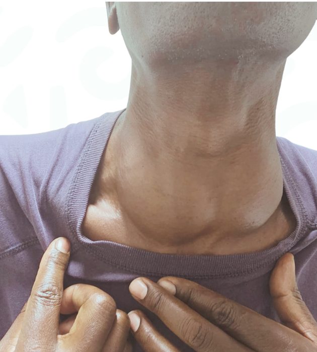
A thyroid nodule is a discrete lump that forms within the thyroid — the butterfly-shaped gland at the front of your neck. Nodules can be solid, fluid-filled (cystic) or mixed; single or multiple. Most grow slowly, cause no symptoms at all, and are discovered by chance on a scan done for something else entirely.
Finding one is rarely an emergency — but every nodule deserves a clear answer to two questions: is it benign, and is it causing (or likely to cause) problems? Those two answers, not fear, should decide what happens next.
Worth knowing
Roughly 19 in every 20 thyroid nodules are benign. The purpose of assessment is to identify the small minority that need more — and to spare everyone else unnecessary procedures.
You are in very ordinary company
1 in 2
By ultrasound, up to half of adults have at least one thyroid nodule by their 60s — most never know it.
4×
Nodules are roughly four times more common in women than in men, and become more frequent with age.
~95%
The overwhelming majority of thyroid nodules prove benign on proper specialist assessment.
Many nodules are found incidentally — on a neck ultrasound, CT scan or carotid study done for another reason. An incidental finding still deserves a structured assessment, but it is very rarely urgent.
What a nodule can be
- Benign colloid nodules — the most common type
- Thyroid cysts — fluid-filled and almost always benign
- Multinodular goitre — several nodules enlarging the gland
- Autoimmune thyroid disease (Hashimoto's thyroiditis)
- Benign thyroid adenomas
- Overactive ('toxic') nodules producing excess hormone
- Thyroid cancer — the least common cause
Iodine levels, family history and previous neck radiation all influence nodule formation. Most of the time, though, no single cause is found — nodules are simply a very common part of how the thyroid ages.
Nodule, goitre — or both?
A goitre is enlargement of the whole gland; a nodule is a lump within it. They often occur together as a nodular goitre, and both are assessed in the same ultrasound examination. Read our goitre guide →
What a nodule can feel like
Common signs
- A lump or swelling you can see or feel at the base of the neck
- A feeling of pressure, fullness or tightness
- Discomfort when swallowing
- A sensation of something in the throat
- Discomfort lying flat or turning the head
- Often — no symptoms at all
If the nodule is overactive
- Palpitations or a racing heart
- Unintended weight loss
- Sweating and heat intolerance
- Anxiety or a fine tremor
- These suggest an overactive ('toxic') nodule
- Confirmed with simple blood tests
See a specialist promptly if you notice
- A nodule growing quickly over weeks
- A hard, fixed lump
- Persistent hoarseness or voice change
- Difficulty swallowing or breathing
- Enlarged lymph nodes in the neck
These signs don't necessarily mean anything serious — but they are the ones that deserve prompt specialist review rather than watchful waiting.
Could my nodule be cancer?
It's the first question almost everyone asks — and the honest answer is reassuring. Only around 5–10% of thyroid nodules prove to be cancerous, and most thyroid cancers found early are among the most treatable of all cancers.
Modern assessment is precise. Ultrasound features graded with ACR TI-RADS, combined with biopsy where indicated, mean we can confidently separate the nodules that need action from the great majority that simply need reassurance or monitoring.
Features that raise attention
- Rapid growth over weeks to months
- A hard, fixed lump
- Persistent hoarseness or voice change
- Enlarged neck lymph nodes
- Previous radiation to the head or neck, especially in childhood
- A family history of thyroid cancer
- A new nodule at a young age, or in men
Features that reassure
- Soft, smooth, mobile nodules
- Purely cystic (fluid-filled) nodules
- Size stable over years
- Benign-pattern ultrasound features (TI-RADS 1–2)
- A benign FNA biopsy result
Six steps to a confident answer
Specialist consultation
Your story, your symptoms and your concerns — heard properly by a consultant endocrinologist.
Examination & blood tests
Careful examination of the neck, plus thyroid function tests to check whether the nodule affects your hormones.
High-resolution ultrasound
Detailed imaging of the nodule and the whole gland — size, composition, features and lymph nodes.
TI-RADS risk grading
Every nodule is graded with the ACR TI-RADS system, so decisions follow evidence rather than habit.
Biopsy only when indicated
Image-guided FNA (or core biopsy) is recommended only when the grading calls for it.
A personalised plan
A clear diagnosis and a plan built around your nodule, your symptoms and your life.
Not all nodules are the same
Nodules are grouped by what they are made of and by whether they are active. Ultrasound tells these apart at your first visit — and the type strongly influences what, if anything, happens next.
Solid nodules
Made up entirely of thyroid tissue. The most common type, and the kind graded most carefully on ultrasound — most are benign, but solid nodules are where TI-RADS features and, when indicated, a biopsy matter most.
Cystic (fluid-filled) nodules
Filled with fluid rather than solid tissue, and almost always benign. If large or uncomfortable they can sometimes be drained in clinic, and cysts that refill respond well to ablation.
Mixed (complex) nodules
Part solid, part fluid — very common. The solid, growing portion is what we assess on ultrasound and, if needed, biopsy; a large fluid component is generally reassuring.
Hyperfunctioning ('hot') nodules
Produce thyroid hormone on their own, which can make the thyroid overactive. Almost always benign — confirmed with blood tests and a radionuclide uptake scan, then managed on their own pathway. More on toxic nodules →
Composition is one of the first things ultrasound measures, and it changes everything: a purely fluid-filled nodule and a solid one with suspicious features are assessed very differently. A benign or malignant label, by contrast, comes only from the full picture — ultrasound features plus, where needed, a biopsy.
What your thyroid bloods add
Every nodule assessment includes simple thyroid blood tests. Most people with a nodule have completely normal thyroid function — but the results tell us whether the nodule is doing anything to your hormones, and they guide the next step.
A normal result is the most common — and most reassuring — finding. Where the numbers are abnormal, they point us to the right test rather than a reflex biopsy.
What the results can flag
- TSH, free T4 and free T3 show whether the nodule is affecting your hormone levels
- A low TSH can signal an overactive ('toxic') nodule — the cue for an uptake scan, not a biopsy
- Calcitonin is checked in selected cases, such as a family history of medullary thyroid cancer
Blood tests measure how the thyroid is working; ultrasound shows how it is built. A complete assessment needs both.
The single most important test
High-resolution ultrasound is the single most important test for a thyroid nodule — painless, radiation-free, and done in the clinic at your visit.
What the scan evaluates
- Nodule size, in three dimensions
- Solid, cystic or mixed composition
- Shape, margins and echogenicity
- Calcifications and other risk features
- Blood-flow pattern
- The rest of the gland and neck lymph nodes
Why it matters
- Same-visit answers — no waiting for reports
- Evidence-based biopsy decisions via TI-RADS
- A baseline for accurate future comparison
- Guidance for FNA, core biopsy and RFA
How we grade a nodule's risk
ACR TI-RADS (Thyroid Imaging Reporting and Data System) scores a nodule's ultrasound features — composition, echogenicity, shape, margins and calcifications — and places it in one of five categories. The category, together with size, tells us whether a biopsy is needed at all.
TR1 — Benign
No suspicious features. No biopsy needed.
TR2 — Not suspicious
Benign-pattern nodule. No biopsy needed.
TR3 — Mildly suspicious
Biopsy considered from about 2.5 cm; ultrasound follow-up from 1.5 cm.
TR4 — Moderately suspicious
Biopsy considered from about 1.5 cm; follow-up from 1 cm.
TR5 — Highly suspicious
Biopsy considered from about 1 cm; close surveillance even when smaller.
The size thresholds are guideline figures — your overall picture decides. What matters is that biopsy decisions follow evidence: graded properly, most nodules never need a needle at all. Specialist interpretation of the scan is what makes the grading trustworthy.
When a nodule needs a closer look
A biopsy answers the one question a scan can't: what the cells themselves are doing. Both types are quick, image-guided, and done in clinic.
Fine-needle aspiration (FNAC)
- A very fine needle samples cells from the nodule under live ultrasound guidance
- Quick — usually minutes, in clinic, with minimal discomfort
- Reported on the standardised Bethesda system, which grades how likely a nodule is to be benign
- For solid nodules, two concordant benign results give the confidence needed before treatment such as RFA
Core needle biopsy (CNB)
- Takes a fine cylinder of tissue rather than cells alone
- Useful when FNA results are repeatedly inconclusive
- Done under local anaesthetic with ultrasound guidance
- Adds certainty in selected nodules before treatment decisions
After either biopsy you can go straight back to your day — a small plaster, perhaps mild bruising, and normal activities the same day.
The Bethesda system, in plain terms
Bethesda I — Non-diagnostic
Too few cells to interpret. The usual answer is simply to repeat the sample.
Bethesda II — Benign
The most common result and a very reassuring one — routine ultrasound monitoring is all that is needed.
Bethesda III — Atypia of undetermined significance
An in-between result; often a repeat sample, molecular testing or a diagnostic operation to settle it.
Bethesda IV — Follicular neoplasm
Usually needs a diagnostic operation (lobectomy), because the cell test alone cannot fully separate benign from malignant.
Bethesda V — Suspicious for malignancy
Surgery is usually recommended, planned with your specialist.
Bethesda VI — Malignant
A clear result that points to definitive treatment — most early thyroid cancers are highly treatable.
Each category carries a different likelihood of cancer, rising from I to VI. Your specialist explains exactly what yours means and what it does — and doesn't — call for. See the full Bethesda system on our FNAC page.
Assessment first — then the right treatment, together
Active surveillance
Most benign nodules need nothing more than scheduled ultrasound checks — no treatment at all.
Medication
Overactive nodules and thyroid hormone problems are managed medically by our endocrinologist.
Aspiration for cysts
Fluid-filled nodules can be drained in clinic; cysts that refill respond well to ablation.
Radiofrequency ablation (RFA)
Benign, symptomatic nodules shrunk without surgery — no incision, no scar, home the same day.
Learn more →
Surgery — selected patients
For cancer, suspicious nodules, or disease genuinely unsuitable for ablation. We refer and guide you.
A decision made with you
Options weighed openly against your symptoms, your scan findings and your preferences — never by default.
Shrinking a nodule without surgery
For benign, biopsy-proven nodules causing a visible swelling, pressure or distress, radiofrequency ablation has changed what treatment means: the nodule is shrunk from within, your thyroid is preserved, and there's no scar.
Who it suits — and who it doesn't
RFA suits benign nodules (usually two concordant benign biopsies for solid nodules) that are causing symptoms, growing, or visibly swelling the neck — and selected overactive nodules and recurrent cysts. It is never used for cancerous or suspicious nodules, and treatment is deferred in pregnancy.
How the treatment works
Under local anaesthetic, a fine electrode is guided into the nodule by ultrasound. Protective fluid (hydrodissection) shields nearby structures where needed, and the nodule is treated in small, precisely controlled units — the moving-shot technique — so energy stays inside the nodule. The treated tissue is then cleared naturally by your body over the following months.
What to expect — and what results look like
You're awake and talking throughout, the ablation itself typically takes 30–60 minutes, and you go home the same day — most people are back to normal within a day or two. Studies report nodule volume roughly halving by six months, with reduction of up to ~90% over longer follow-up; mild soreness or temporary swelling can occur, and serious complications are uncommon.
Follow-up
Ultrasound at around one, three-to-six and twelve months tracks the shrinkage — coordinated remotely for patients who travel to us from across East Africa.
Sometimes an operation is the right call
Surgery remains the right treatment in specific situations: a nodule confirmed or suspicious for cancer, a very large or retrosternal goitre causing significant compression, or disease that genuinely isn't suitable for ablation.
When surgery is the answer, we say so plainly — and coordinate your referral to the right surgical team, staying involved in your thyroid care before and after the operation. The decision is always shared, never rushed.
Our promise
You'll never be steered toward a treatment because it's the one we offer. Benign and symptomatic — we'll discuss RFA. Suspicious or malignant — we'll get you to the right surgeon, quickly.
Follow-up that doesn't drop you
Benign nodules on surveillance
Low-risk nodules are typically re-scanned at 12–24 month intervals, spacing out as stability is confirmed. Many need nothing more, ever.
After RFA
Ultrasound at around 1, 3–6 and 12 months tracks the volume reduction — with remote coordination for international patients.
After surgery
Wound review, calcium and thyroid hormone checks, and long-term thyroid function monitoring with replacement adjusted as needed.
Your thyroid function
Thyroid blood tests are repeated when symptoms or findings indicate — not reflexively. Long-term continuity is the point: one clinic, one record, one specialist who knows your neck.
Thyroid nodules — your questions answered
Very. By ultrasound, up to half of adults have at least one nodule by later life, and most never cause any problem. Being told you have a nodule is common; what matters is assessing it properly.
Only around 5–10% of nodules prove cancerous, and most thyroid cancers found early are very treatable. Ultrasound grading with TI-RADS, plus a biopsy when indicated, tells the two apart confidently.
No — most don't. We grade every nodule with ACR TI-RADS; a biopsy is only recommended when the nodule's features and size cross evidence-based thresholds.
Very little. FNA uses a needle finer than the one used for blood tests, takes minutes under local anaesthetic if needed, and you return to normal activities the same day.
Cystic (fluid-filled) nodules can shrink or resolve, and some small nodules stay unchanged for life. Solid nodules rarely disappear, which is why surveillance or treatment is planned around what yours is doing.
Many stay stable for years. Some grow slowly, and a minority grow enough to cause pressure or visible swelling — that's when treatment such as RFA becomes worth discussing.
Usually not — most patients with nodules have normal thyroid function. An overactive ('toxic') nodule can cause hyperthyroidism, which is why thyroid blood tests are part of every assessment.
Yes, in almost all cases. A benign nodule doesn't restrict exercise, flying or daily life. If you have symptoms of an overactive thyroid, get that controlled first.
Nodules found in pregnancy are assessed with ultrasound as normal — it's completely safe. Biopsy can be done in pregnancy where needed; treatments like RFA are simply deferred until after delivery.
Often, yes. Benign, biopsy-proven nodules causing symptoms can usually be shrunk with radiofrequency ablation — no incision, no scar, home the same day, thyroid preserved.
After RFA, almost never — your healthy thyroid is preserved. After surgery that removes part or all of the gland, hormone replacement is often needed lifelong.
It depends on its TI-RADS grade and history — typically ultrasound at 12–24 month intervals for benign nodules, spacing out as stability is confirmed. Your specialist sets the right interval for you.
Care built around your thyroid
- Consultant endocrinologist with dedicated thyroidology expertise
- In-clinic high-resolution thyroid ultrasound at your first visit
- Image-guided FNA and core needle biopsy
- Kenya's only centre offering thyroid RFA
- Evidence-based care aligned with ATA, ETA and BTA guidance
- Multidisciplinary referral network for surgical and cancer care
Meet your specialist
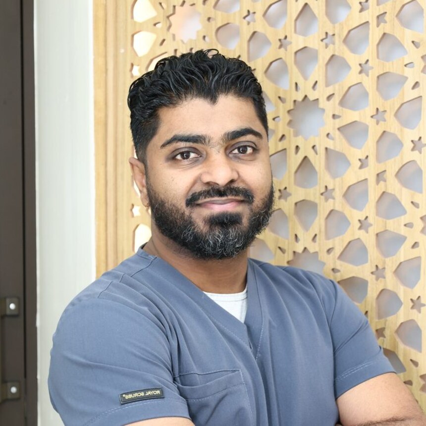
Consultant Physician · Interventional Endocrinologist & Diabetologist — one of the first doctors in Kenya to offer thermal ablation of thyroid nodules.
From first message to long-term peace of mind
Book
Message us on WhatsApp or book online — with or without previous scans.
Consult
An unhurried consultation about your nodule, symptoms and history.
Scan
High-resolution ultrasound with TI-RADS grading, at the same visit.
Investigate
Blood tests, and a biopsy only if the grading calls for one.
Decide
A shared decision: surveillance, medication, RFA or referral.
Treat
Treatment arranged promptly — often as a single day-case.
Follow up
Scheduled reviews for as long as your thyroid needs them.
Medically reviewed by Dr. Ebrahim Yusuf Ebrahim, Consultant Physician & Interventional Endocrinologist. Clinical figures are ranges from peer-reviewed studies, provided for general education — individual results vary and are confirmed at your review.
A nodule deserves an answer — not months of worry
Book a specialist assessment, or send your existing ultrasound and biopsy on WhatsApp for a reports review. Either way, you'll know where you stand.